Auxetic Scaffolds for Tissue Engineering
The global tissue engineering market is worth ~$2.59 to $11 billion (2012-2014), with significant growth forecast to reach $27 billion by 2018 and $73 billion by 2025). The major market focus is related to orthopaedics, skin, oncology, cardiovascular, dental, neurology and urinary systems. Tissue engineering scaffolds for regenerative therapies are required to match target tissue attributes, withstand a variety of loading conditions, support and stimulate cell growth, and maintain the normal anatomy and physiology of the organ.
Natural biological tissues are known to display auxetic characteristics, the material becomes thicker, rather than thinner, when stretched; corresponding to negative Poisson’s ratio. These properties have been reported in skin, artery, tendon, embryonic tissues, and cancellous bone. Mechanical stimulation of the scaffold promotes cell proliferation and a recent study indicated enhanced proliferation may occur in an auxetic scaffold. Auxetic porous materials facilitate mass transport and, therefore, there is potential for optimal flow or delivery of nutrients, metabolic wastes and therapeutic agents. For these reasons, the development of porous auxetic materials promises to deliver improved next-generation tissue engineering scaffolds.
This project is a collaboration between the Biomolecular Sciences Research Centre (BMRC) and MERI, initially funded by SCH, with further funding via a SHU VC PhD scholarship matched by an external biomedical devices company. The overarching aim of the project is to develop a range of ‘auxetic’ porous scaffolds for eventual tissue engineering applications, which mimic the mechanical properties of soft tissues, promote migration, adhesion and differentiation of cells, and enable delivery of bioactive components. The produced scaffolds are being characterised for their structure and mechanical properties, prior to investigation in vitro. Tissue cells are being cultured on the scaffolds and a variety of biochemical, histological, immunohistological, molecular biology, scanning electron microscopy and biomechanical analysis methods are being used to determine the cell behaviour and interaction with the auxetic scaffolds (Figure).
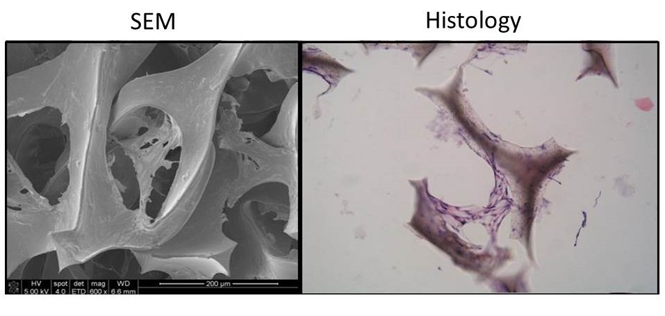 Figure. Cells and extracellular matrix forming bridges within auxetic foam scaffold pores